
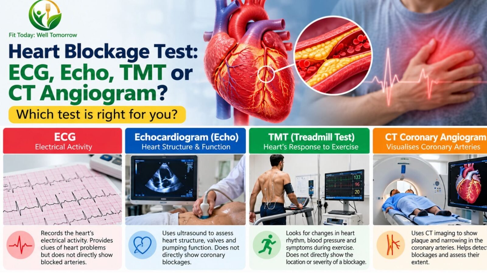
If you are looking for a heart blockage test, the one current guidelines put first is a CT coronary angiogram. For new chest pain of suspected cardiac origin, NICE guideline CG95 offers it as the primary test and the 2024 European Society of Cardiology guidelines give it a Class I, Level A recommendation, because it shows directly whether the coronary arteries are narrowed or blocked.
An ECG and an echocardiogram are still done early, but they answer different questions: the ECG looks at electrical rhythm and damage, the echo at pumping function, valves and muscle.
A treadmill test (TMT) is a weak tool for diagnosing blocked arteries. It is better used to judge fitness and risk than to rule disease in or out.
That ordering may be unfamiliar if you have been sent straight for a treadmill test. Here is what each test measures, what it cannot see, and who needs which one.
The four tests, side by side
| Test | What it measures | Radiation | Good at | Poor at |
|---|---|---|---|---|
| ECG (electrocardiogram) | Electrical activity and rhythm | None | Arrhythmias, evidence of a heart attack, conduction problems | Ruling out angina or narrowed arteries |
| Echocardiogram (echo) | Structure and pumping, using ultrasound | None | Valve disease, heart failure, cardiomyopathy, ejection fraction | Showing whether coronary arteries are blocked |
| TMT (exercise ECG) | ECG changes and symptoms during exercise | None | Exercise capacity, risk stratification | Diagnosing obstructive coronary disease |
| CT coronary angiogram | Anatomy of the coronary arteries | Yes | Ruling out obstructive coronary disease | Judging whether a borderline narrowing limits blood flow |
What an ECG does, and what it misses
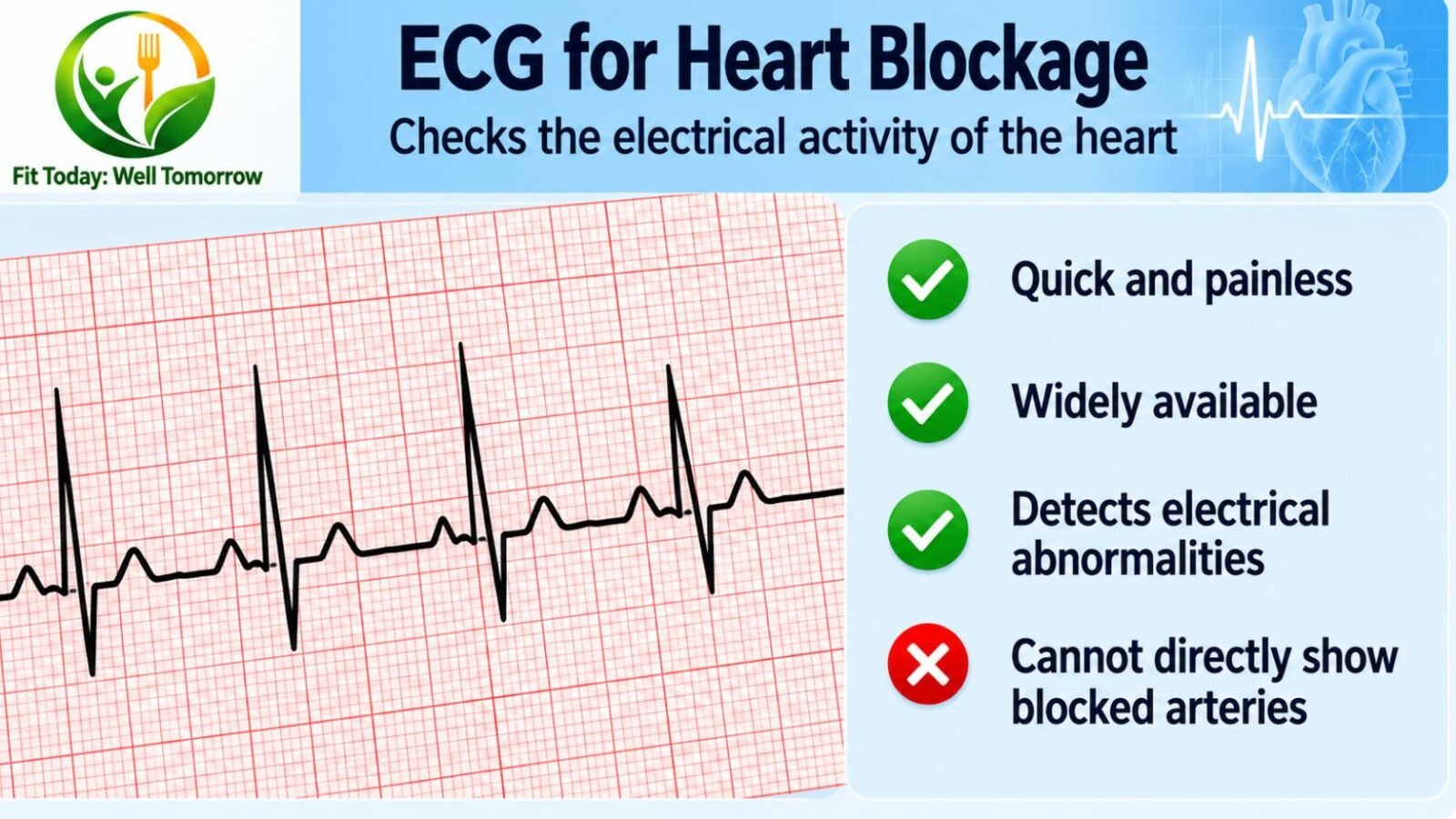
An ECG checks the heart’s rhythm and electrical activity, using sensors stuck to the skin that pick up the electrical signal the heart produces with each beat, according to NHS Inform. It takes a few minutes, is painless, and no electricity is put into your body.
It can identify arrhythmias where the heart beats too slowly, too quickly or irregularly, and it can show evidence of coronary heart disease, heart attacks and cardiomyopathy. There are three main versions: a resting ECG lying down, a stress or exercise ECG on a treadmill or bike, and an ambulatory ECG worn at the waist for a day or more.
Here is the part that gets lost. A normal ECG does not mean your coronary arteries are clear. NICE CG95, as reproduced by the clinician reference GPnotebook, is blunt about it: do not rule out a diagnosis of stable angina on the basis of a normal resting 12-lead ECG. NHS Inform makes the same point more gently, noting that other tests may also need to be carried out before it is possible to tell you whether there is a problem.
The 2024 European Society of Cardiology guidelines on chronic coronary syndromes place the resting 12-lead ECG, alongside transthoracic echocardiography, as a Class I recommendation in initial evaluation, though at Level of Evidence C, and for specific reasons: to assess cardiac function, detect alternative diagnoses, and risk stratify. Not to diagnose blocked arteries. The ECG is a starting point, not a verdict.
What an echocardiogram shows
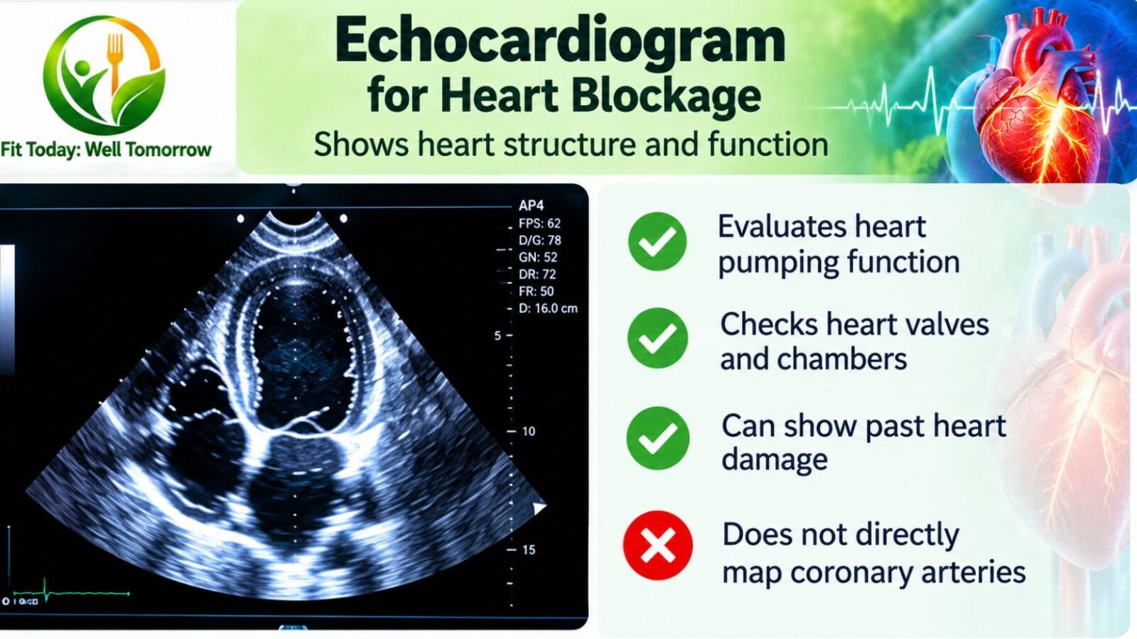
An echocardiogram is an ultrasound scan used to look at the heart and nearby blood vessels. No radiation is involved, so there is no cumulative dose to weigh if it needs repeating, and the whole procedure usually takes between 15 and 60 minutes.
It is the right test when the question is about the heart as a pump or a structure. NHS 111 Wales patient information lists damage from a heart attack, heart failure, congenital heart disease, valve problems, cardiomyopathy and endocarditis among the conditions it helps detect. Variants include the standard transthoracic scan, a transoesophageal scan where a small probe is passed down the oesophagus, a stress echocardiogram, and a contrast echocardiogram.
The number most people take away from an echo report is ejection fraction, the percentage of blood the left ventricle pushes out with each beat. Cleveland Clinic puts the normal range at 50% to 70%, with 40% to 49% counted as mildly reduced and 39% or less classified as heart failure with reduced ejection fraction. The echocardiogram is the most common way ejection fraction is measured.
If your main complaint is breathlessness or swollen ankles rather than chest pain, this is usually the test that matters. In the UK, NICE guideline NG106 routes suspected heart failure through a blood test first: an NT-proBNP above 2,000 ng/l warrants specialist review within two weeks, and a result between 400 and 2,000 ng/l within six weeks, with that specialist assessment including a transthoracic echocardiogram.
What an echo will not tell you is whether a particular coronary artery is narrowed. The ESC’s stated reasons for ordering a transthoracic echocardiogram early are to assess cardiac function, detect alternative diagnoses and risk stratify, not to inspect the coronary arteries.
TMT: why the treadmill test has been demoted
This is the finding most worth knowing, because the treadmill test is often the first thing people are sent for.
The 2024 ESC guidelines state it plainly: due to its low sensitivity (58%) and specificity (62%), exercise ECG testing has low diagnostic performance for the diagnosis of obstructive CAD and should mainly be used for risk stratification. Exercise ECG now carries only a Class IIb, Level B recommendation, and for a narrow purpose: it should be considered in patients with low clinical likelihood of obstructive CAD to reclassify to very low likelihood.
A 2025 systematic review in the Journal of Clinical Medicine pooled 104 studies covering 16,824 symptomatic people with suspected or known coronary disease, and found the same hierarchy. Exercise stress testing had a sensitivity of 0.66 (95% CI 0.59 to 0.72) and a specificity of 0.61 (0.55 to 0.67).
Stress echocardiography reached 0.81 and 0.85. Stress SPECT reached 0.82 and 0.74. Stress cardiac MRI topped the list at 0.83 and 0.89. The authors concluded that exercise stress testing demonstrated the lowest accuracy for detecting obstructive CAD, whereas stress CMR exhibited the highest.
Read the sensitivity figure carefully. A sensitivity around 0.66 means that of every three people who do have obstructive coronary disease, roughly one will have a treadmill test that fails to flag it. A specificity around 0.61 means false alarms are common too, which is how people end up with an invasive angiogram they did not need.
In fairness to the review’s own caveats, heterogeneity across the included studies was high (I² between 82.5% and 92.5%) and the authors found statistically significant publication bias for all four methods. These are pooled estimates with wide real-world scatter, not precision instruments.
Outcomes data points the same way. A 2023 systematic review in the Annals of Internal Medicine found that CT coronary angiography was associated with a reduction in cardiovascular death and myocardial infarction compared with exercise ECG, with a relative risk of 0.66 (95% CI 0.44 to 0.99) at moderate certainty, and compared with SPECT perfusion imaging at a relative risk of 0.64 (0.45 to 0.90) at high certainty.
None of this makes a TMT useless. The ESC’s own verdict is that it should mainly be used for risk stratification, and it is the one test here that shows what you can actually do under load. It is a fitness and risk test. It is a poor gatekeeper for the catheterisation lab.
CT coronary angiogram: now the recommended first test
A CT coronary angiogram uses a type of X-ray called computed tomography, with dye injected into a vein, to show whether the coronary arteries are narrowed or blocked. No catheter goes anywhere near your heart.
NICE’s plain-English guidance for patients says people with suspected stable angina should be offered CT coronary angiography as the primary test. That was a deliberate shift.
As a 2017 review in Current Cardiovascular Imaging Reports describes it, the updated 2016 NICE guideline is notable for its removal of the pre-test probability model and the use of CTCA as the first-line investigation in all patients with atypical or typical angina symptoms, or those who are asymptomatic with ECG changes suggesting ischaemia. The older version sorted patients by estimated likelihood: calcium scoring at 10% to 29%, functional imaging at 30% to 60%, invasive angiography at 61% to 90%. That ladder is gone.
The ESC agrees, giving CCTA as a first-line diagnostic test a Class I, Level A recommendation, with non-invasive stress imaging recommended where CT is unavailable or inconclusive, also Class I, Level A.
The accuracy data behind that is solid. An individual patient data meta-analysis in the BMJ in 2019 pooled 5,332 patients from 65 prospective studies and found a sensitivity of 95.2% (92.6 to 96.9) and a specificity of 79.2% (74.9 to 82.9).
That gap between the two numbers is the whole story of how to use the test. At a 7% pre-test probability, the negative predictive value was 97.8%, while the positive predictive value was only 50.9%. In plain terms: a clean CT scan is very reassuring, and an abnormal one often needs something else to confirm it.
At a higher pre-test probability of 67%, the positive predictive value rose to 82.7%. Across a pre-test probability range of 7% to 67%, the analysis found that CT could move a patient past a treat threshold (post-test probability above 50%) or a no-treat threshold (below 15%). That is why a doctor should assess you before ordering the scan rather than after.
Performance is not uniform. The same analysis found accuracy slightly lower in women than in men, and lowest in people older than 75.
Where CT scans struggle
High or irregular heart rates and heavy calcification all reduce the diagnostic accuracy of the technique, as a 2010 British Journal of Cardiology review puts it. Heavy coronary calcification is likewise described in Acta Radiologica as a major limitation of the scan.
The scan is best performed in those in sinus rhythm, and beta blockers and sublingual nitrates are routinely administered to slow the heart and open the arteries before scanning.
That is worth knowing before you arrive. If you have atrial fibrillation, a fast resting pulse, or extensive calcium from years of untreated risk factors, the scan may be harder to read, and your cardiologist may reasonably choose a functional test instead.
What about the radiation?
It has fallen sharply. A worldwide registry of 4,502 patients across 61 hospitals in 32 countries found the median dose-length product for cardiac CT angiography in 2017 was 195 mGy·cm (interquartile range 110 to 338), a 78% reduction compared with 2007. Non-diagnostic scan rates barely moved, 1.7% in 2007 against 1.9% in 2017.
Dose is not zero, and it is a real consideration in younger patients. But that registry compared 2017 with 2007, so the scan protocols in use now are a long way from the ones the early dose warnings were written about.
For comparison, the Cardiac Society of Australia and New Zealand puts the radiation exposure of a coronary calcium scan at about 0.5 to 1.0 mSv, similar to breast mammography. An older British Journal of Cardiology review put it at 1 to 2 mSv, so treat that as a range rather than a single figure.
Invasive coronary angiography: still the reference, no longer the default
Invasive coronary angiography involves a fine, hollow tube called a catheter being inserted through the artery of the person’s leg or arm and up to the heart, with contrast and X-rays used to map the arteries. It is the test the others in this article were measured against in the diagnostic-accuracy studies quoted here.
What changed is who should get it first. The DISCHARGE trial randomised 3,561 patients with stable chest pain, 56.2% of them women, to CT or straight to invasive angiography. Major adverse cardiovascular events occurred in 2.1% of the CT group against 3.0% of the invasive group, a difference that did not reach statistical significance (hazard ratio 0.70; 95% CI 0.46 to 1.07; P=0.10).
The complication figures were the clearer result. Major procedure-related complications occurred in 0.5% of the CT group against 1.9% of those sent for invasive angiography, a hazard ratio of 0.26 (95% CI 0.13 to 0.55). Roughly a four-fold difference in procedural harm, for no measurable gain in event prevention.
SCOT-HEART, which followed 4,146 patients with stable chest pain for five years, adds the other half of the picture. Coronary heart disease death or non-fatal myocardial infarction occurred in 2.3% of those who had a CT scan added to standard care, against 3.9% with standard care alone (hazard ratio 0.59; 95% CI 0.41 to 0.84; P=0.004). Rates of invasive angiography (491 against 502) and revascularisation (279 against 267) were essentially identical between the groups.
So the benefit did not come from more stenting. Patients in the CT arm started more preventive and anti-anginal treatment, with no difference in overall mortality. The scan changed what doctors prescribed, not how often they intervened mechanically.
Where coronary calcium scoring fits
A calcium score is a CT scan of the coronary arteries measured without contrast, reported as an Agatston score. It is a risk tool, not a diagnostic one.
The Cardiac Society of Australia and New Zealand position statement recommends it for individuals without coronary disease aged 45 to 75 years with an absolute five-year cardiovascular risk of 10% to 15% who are asymptomatic. It explicitly does not recommend it for patients who have symptomatic or previously documented coronary artery disease. If you have chest pain, this is not your test.
The score bands are: 0 very low risk, 1 to 99 low, 100 to 399 intermediate, and 400 or above, or above the 75th percentile, high. A score of zero indicates no detectable coronary atherosclerosis, with a very low event rate, which is why it is useful for deciding whether a borderline-risk person needs a statin.
Which test, when
New chest or arm discomfort that comes on with exertion and eases with rest. See a doctor promptly. Expect a resting ECG and usually an echo as part of initial assessment, then a CT coronary angiogram as the test that settles the question. A normal ECG does not close the case.
Breathlessness, ankle swelling, or reduced exercise tolerance without chest pain. The echocardiogram is the central test, often after a natriuretic peptide blood test. You are looking for a pump or valve problem, not a blockage.
Palpitations, skipped beats, dizziness or blackouts. A resting ECG is a start, but if symptoms come and go, an ambulatory ECG monitors you at home for one or more days, so it can record an episode a few minutes of resting trace would miss.
Known coronary disease with new or worsening symptoms. This is a conversation about functional testing or invasive angiography with your cardiologist, not a reason to self-refer for a scan. The American College of Cardiology’s summary of the ESC guideline frames the choice as resting on pre-test likelihood, other patient characteristics that influence test performance, and local expertise and availability.
No symptoms at all, just worried. See the next section.
Should you have these tests if you have no symptoms?
Probably not, and a routine heart test in a health package is one of the easier ways to spend money without benefit.
The US Preventive Services Task Force recommends against screening with resting or exercise electrocardiography to prevent cardiovascular disease events in asymptomatic adults at low risk of CVD events, where low risk means a 10-year event risk below 10%. For asymptomatic adults at intermediate or high risk, it concluded that the current evidence is insufficient to assess the balance of benefits and harms.
Insufficient is not the same as beneficial. If you have no symptoms, a routine ECG or TMT bundled into a health package is unlikely to help you and can start a chain of follow-up tests, each with its own cost and risk.
What does help is knowing and treating your risk factors. Cardiovascular disease accounted for 28.1% of all deaths in India in 2016, up from 15.2% in 1990, with 23.8 million people living with ischaemic heart disease, a 2.3-fold rise over the same period.
More than half of cardiovascular deaths in India that year, 53.4%, were in people younger than 70. Blood pressure, lipids, blood sugar, smoking and weight are where that burden is decided, not in the scan queue.
The CSANZ position statement supports calcium scoring in asymptomatic people aged 45 to 75 whose absolute five-year cardiovascular risk is 10% to 15%, which is the one asymptomatic use here with a named guideline behind it. Whether it should change a statin decision in your case is a question for your doctor.
What these tests cost in India
Government schedules are the most reliable public benchmark, since private pricing varies enormously by city and hospital. The following are reimbursement rates, not quotes, and the higher figure in each pair applies to NABH-accredited centres.
| Test | NABH rate | Non-NABH rate | Source |
|---|---|---|---|
| ECG | ₹175 | ₹150 | CGHS OM, 15/06/2023 |
| Echocardiography | ₹1,475 | ₹1,255 | CGHS OM, 15/06/2023 |
| Treadmill test (TMT) | ₹1,120 | ₹950 | Delhi Arogya Kosh, 31/08/2023 |
| Stress echocardiography | ₹2,400 | ₹2,040 | CGHS OM, 15/06/2023 |
| Dobutamine stress echo | ₹3,000 | ₹2,550 | CGHS OM, 15/06/2023 |
| Coronary angiography (invasive) | ₹13,225 | ₹11,240 | CGHS OM, 19/12/2023 |
One honest gap: none of the three official rate lists checked here carries a rate for CT coronary angiography, so there is no government benchmark to quote for the test that guidelines now put first. Ask for a written estimate before booking, and ask whether contrast and the pre-scan medication are included.
The ordering is worth noting. An ECG is reimbursed at ₹150 to ₹175. An invasive angiogram is reimbursed at roughly seventy-five times the higher of those, and in the DISCHARGE trial major procedure-related complications occurred in 1.9% of the patients sent for one. The cheap test is cheap partly because it answers a narrow question.
Frequently asked questions
Can an ECG detect a blocked artery?
Not reliably. An ECG can show evidence of coronary heart disease or a heart attack, but NICE CG95, as reproduced by GPnotebook, specifically says not to rule out stable angina on the basis of a normal resting 12-lead ECG. A clear ECG is reassuring about rhythm, not about your arteries.
Is a TMT enough to rule out heart disease?
No. The ESC puts exercise ECG sensitivity at 58% and specificity at 62% for diagnosing obstructive coronary disease, and a 2025 pooled analysis of 104 studies found sensitivity of 0.66 and specificity of 0.61. Roughly one in three people with real disease will pass a treadmill test. It is better used to assess exercise capacity and risk.
Which is better, a TMT or a CT coronary angiogram?
For diagnosing whether an artery is narrowed, the CT scan, clearly. Pooled CT accuracy is 95.2% sensitivity and 79.2% specificity, and a 2023 review found CT associated with fewer cardiovascular deaths and heart attacks than exercise ECG, at a relative risk of 0.66 (95% CI 0.44 to 0.99), rated moderate certainty. The treadmill test retains value for judging fitness and prognosis.
Do I need an echo and a CT angiogram, or just one?
Often both, because they answer different questions. The ESC recommends transthoracic echocardiography in initial evaluation, at Class I but Level of Evidence C, to assess cardiac function, find alternative diagnoses and risk stratify. The CT answers whether the coronary arteries are obstructed. Neither substitutes for the other.
Is a CT coronary angiogram safe?
It involves X-ray radiation and iodinated contrast, so it is not free of risk, but dose has dropped substantially: a worldwide registry found the median dose-length product in 2017 was 78% lower than in 2007. Compared with invasive angiography, major procedure-related complications in the DISCHARGE trial were 0.5% with CT against 1.9% with the invasive route.
What if my calcium score is zero?
A score of zero indicates no detectable coronary atherosclerosis, with a very low event rate. That is genuinely good news for risk prediction in an asymptomatic person. It is not a reason to ignore new chest pain, and calcium scoring is explicitly not recommended for people who already have symptoms.
Why did my doctor give me a tablet before my CT scan?
Because image quality depends on a slow, regular heartbeat. Beta blockers and sublingual nitrates are routinely administered before CT coronary angiography, since high or irregular heart rates reduce the accuracy of the scan.
I feel fine. Should I add an ECG or TMT to my annual health check?
The USPSTF recommends against screening with resting or exercise ECG in asymptomatic adults at low cardiovascular risk, and found the evidence insufficient at intermediate or high risk. Money and attention are better spent on blood pressure, lipids, blood sugar and smoking.
Which test is best for a woman with chest pain?
The same pathway applies, with one caveat worth raising with your doctor: the BMJ meta-analysis found CT accuracy slightly lower in women than in men. That is a reason for careful clinical assessment alongside the scan, not a reason to skip it. Notably, 56.2% of the 3,561 patients in the DISCHARGE trial were women.
Before you book anything
Which test you need depends on what your symptoms are, how likely coronary disease is before any scanning happens, and what your local hospital does well. All three of those are judgements a doctor makes with you, which is why the ESC guideline ties test choice to pre-test likelihood, patient characteristics and local expertise and availability rather than to a fixed sequence.
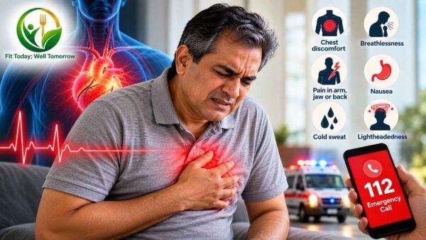
If you have new chest pain, particularly with exertion, do not wait for a convenient appointment slot. If you have crushing central chest pain now, with sweating, nausea or pain spreading to the arm or jaw, treat it as an emergency.
This article explains what the tests measure. It is not a substitute for assessment by your own doctor, who can weigh your symptoms, risk factors and history before deciding which one you need.
Sources
- India State-Level Disease Burden Initiative CVD Collaborators. The changing patterns of cardiovascular diseases and their risk factors in the states of India: the Global Burden of Disease Study 1990-2016. The Lancet Global Health, 2018.
- Vrints C, Andreotti F, Koskinas KC, et al. 2024 ESC Guidelines for the management of chronic coronary syndromes. European Heart Journal, 2024;45(36):3415-3537.
- European Society of Cardiology. New Chronic Coronary Syndrome guidelines expand diagnostic tools. ESC press release, 2024.
- American College of Cardiology. 2024 ESC Guidelines for Management of Chronic Coronary Syndromes: Key Points. 2024.
- National Institute for Health and Care Excellence. Recent-onset chest pain of suspected cardiac origin: investigations for stable angina (information for the public). Clinical guideline CG95.
- National Institute for Health and Care Excellence. Chronic heart failure: diagnosis visual summary. NICE guideline NG106.
- Moss AJ, Dweck MR, Williams MC, Newby DE. The Updated NICE Guidelines: Cardiac CT as the First-Line Test for Coronary Artery Disease. Current Cardiovascular Imaging Reports, 2017.
- Haase R, Schlattmann P, Gueret P, et al. Diagnosis of obstructive coronary artery disease using computed tomography angiography in patients with stable chest pain depending on clinical probability and in clinically important subgroups: meta-analysis of individual patient data. BMJ, 2019;365:l1945.
- Sonaglioni A, Polymeropoulos A, Baravelli M, Nicolosi GL, Lombardo M, Biondi-Zoccai G. Diagnostic Accuracy of Exercise Stress Testing, Stress Echocardiography, Myocardial Scintigraphy, and Cardiac Magnetic Resonance for Obstructive Coronary Artery Disease. Journal of Clinical Medicine, 2025;14(17):6238.
- Zito A, Princi G, Lombardi M, et al. Diagnostic Strategies for the Assessment of Suspected Stable Coronary Artery Disease. Annals of Internal Medicine, 2023.
- DISCHARGE Trial Group. CT or Invasive Coronary Angiography in Stable Chest Pain. New England Journal of Medicine, 2022.
- SCOT-HEART Investigators. Coronary CT Angiography and 5-Year Risk of Myocardial Infarction. New England Journal of Medicine, 2018.
- Stocker TJ, Deseive S, Leipsic J, et al. Reduction in radiation exposure in cardiovascular computed tomography imaging: results from the PROTECTION VI registry. European Heart Journal, 2018;39(41):3715-3723.
- Hamilton-Craig CR, Chow CK, Younger JF, et al. Cardiac Society of Australia and New Zealand position statement executive summary: coronary artery calcium scoring. Medical Journal of Australia, 2017;207(8):357-361.
- US Preventive Services Task Force. Cardiovascular Disease Risk: Screening With Electrocardiography, final recommendation statement. 2018.
- NHS Inform Scotland. Electrocardiogram (ECG). Last updated 07/07/2025.
- NHS 111 Wales. Echocardiogram. Reviewed 07/07/2023.
- Cleveland Clinic. Ejection Fraction. Last updated 14/04/2026.
- Obaid DR, Murray SW, Palmer ND, Rudd JHF. Contemporary coronary imaging from patient to plaque: part 3, cardiac computed tomography. British Journal of Cardiology, 2010;17:235-9.
- Engel LC, Thai WE, Medina-Zuluaga HA, et al. Non-diagnostic coronary artery calcification and stenosis: a correlation of coronary computed tomography angiography and invasive coronary angiography. Acta Radiologica, 2017.
- GPnotebook. Investigation of possible angina (reproducing NICE CG95 recommendation wording). Registration required.
- Central Government Health Scheme, Ministry of Health and Family Welfare. Revision of CGHS rates for 36 radiological and imaging investigations, Office Memorandum dated 15/06/2023.
- Central Government Health Scheme. Revision of CGHS rates for cardiology procedures, F No Z15025/32/2023/DIR/CGHS, dated 19/12/2023.
- Directorate General of Health Services, Government of NCT of Delhi. Delhi Arogya Kosh rate list, Office Memorandum dated 31/08/2023.